MANILA – The World Health Organization (WHO) solidarity trials on the coronavirus disease 2019 (Covid-19) vaccines might start by the last week of October, a health official said Friday.

"(In) the fourth week of October, the implementation might start already. And I say the implementation might start because there could be a lot of factors, intervening factors, that the dates might be moved. But based on the discussions September 9, this is what we have agreed upon," Department of Health Undersecretary Maria Rosario Vergeire said in a mix of Filipino and English during a virtual media forum.

Citing the timeline from the WHO, Vergeire said the vaccines to be included in the solidarity trial would be identified in the third or fourth week of September while the sites for the conduct of the trials would be identified by the end of September.

"We will be monitoring this and we hope we can pursue this based on the specific timeline that was given to us," she said.

The solidarity trial is an international clinical trial to test the effectiveness of a possible vaccine for Covid-19.

The Philippines’ participation in the WHO solidarity trial was approved by the Single Joint Research Ethics Board on April 17 in support of the Covid-19 global response. (PNA-By Ma. Teresa Montemayor)

Source:https://www.pna.gov.ph/articles/1115146 

An antibody test for the virus that causes COVID-19, developed by researchers at The University of Texas at Austin in collaboration with Houston Methodist and other institutions, is more accurate and can handle a much larger number of donor samples at lower overall cost than standard antibody tests currently in use. In the near term, the test can be used to accurately identify the best donors for convalescent plasma therapy and measure how well candidate vaccines and other therapies elicit an immune response.

Additional uses coming later that are likely to have the biggest societal impact, the researchers say, are to assess relative immunity in those previously infected by the SARS-CoV-2 virus and identify asymptomatic individuals with high levels of neutralizing antibodies against the virus.

The UT Austin research team, led by Jason Lavinder, a research associate in the Cockrell School of Engineering, and Greg Ippolito, assistant professor in the College of Natural Sciences and Dell Medical School, developed the new antibody test for SARS-CoV-2 and provided the viral antigens for this study via their UT Austin colleague and collaborator, associate professor Jason McLellan. Other UT Austin team members are Dalton Towers and Jimmy Gollihar. The work was published this week in The Journal of Clinical Investigation.

"This is potentially game-changing when it comes to serological testing for COVID-19 immunity," Lavinder said. "We can now use highly scalable, automated testing to examine antibody-based immunity to COVID-19 for hundreds of donors in a single run. With increased levels of automation, limited capacity for serological testing can be rapidly addressed using this approach."

The gold standard of COVID-19 antibody testing measures the amount of virus-neutralizing (VN) antibodies circulating in the blood because this closely correlates with immunity. However, this kind of antibody testing is not widely available because it's technically complex; requires days to set up, run, and interpret; and needs to be performed in a biosafety level 3 laboratory.

The research team, therefore, looked to another type of test, called ELISA assays, that can be implemented and performed with relative ease in a high-throughput fashion and is widely available and extensively used in clinical labs across the world. The ELISA tests, or enzyme-linked immunosorbent assays, look at whether antibodies against specific SARS-CoV-2 proteins are present and produce a quantitative measure of those antibodies.

The goal of the study was to test the hypothesis that levels of antibodies that target two regions of the virus's spike protein -- spike ectodomain (ECD) and receptor-binding domain (RBD) -- are correlated with virus-neutralizing antibody levels, making these more accessible, easier-to-perform ELISA tests a surrogate marker to identify plasma donors with antibody levels above the recommended U.S. Food and Drug Administration threshold for convalescent plasma donation.

In collaboration with UT Austin, Penn State University, and the U.S. Army Medical Research Institute of Infectious Diseases, study authors James M. Musser, M.D., Ph.D., and Eric Salazar, M.D., Ph.D., physician-scientists at Houston Methodist, used the new test to evaluate 2,814 blood samples used in an ongoing study of convalescent plasma therapy. Houston Methodist became the first academic medical center in the nation to transfuse plasma from recovered individuals into COVID-19 patients.

The researchers found that the ELISA tests had an 80% probability or greater of comparable antibody level to VN levels at or above the FDA-recommended levels for COVID-19 convalescent plasma. These results affirm that all three types of tests could potentially serve as a quantitative target for therapeutic and prophylactic treatments.

Ultimately, the study successfully concluded that anti-RBD or anti-ECD antibody levels can serve as a surrogate for VN levels to identify suitable plasma donors and that these alternate ELISA tests may provide critical information about COVID-19 immunity.

"This research required a perfect storm at the university, which included the extraordinary combination of a world-famous coronavirus structural biology lab, a nimble and passionate visiting army scientist, and the highest echelons of the university's administration who were committed to bringing our extensive research programs to bear on the COVID-19 crisis," Ippolito said.

This study was supported by funding from the National Institutes of Health, the Fondren Foundation, the National Institute of Allergy and Infectious Diseases, the Army Research Office, Houston Methodist Hospital, Houston Methodist Infectious Diseases Research Fund, Houston Methodist Research Institute, and seed funding from the Huck Institutes of the Life Sciences for the studies at Penn State, together with the Huck Distinguished Chair in Global Health award. Funding was also provided through the CARES Act, with programmatic oversight from the Military Infectious Diseases Research Program.


Story Source:

Materials provided by University of Texas at AustinNote: Content may be edited for style and length.


Journal Reference:

  1. Eric Salazar, Suresh V. Kuchipudi, Paul A. Christensen, Todd Eagar, Xin Yi, Picheng Zhao, Zhicheng Jin, S. Wesley Long, Randall J. Olsen, Jian Chen, Brian Castillo, Christopher Leveque, Dalton Towers, Jason J. Lavinder, Jimmy Gollihar, Jose A. Cardona, Gregory C. Ippolito, Ruth H. Nissly, Ian Bird, Denver Greenawalt, Randall M. Rossi, Abhinay Gontu, Sreenidhi Srinivasan, Indira Poojary, Isabella M. Cattadori, Peter Hudson, Nicole M. Josleyn, Laura Prugar, Kathleen E. Huie, Andrew S. Herbert, David W. Bernard, John M. Dye, Vivek Kapur, James M. Musser. Convalescent plasma anti-SARS-CoV-2 spike protein ectodomain and receptor binding domain IgG correlate with virus neutralizationJournal of Clinical Investigation, 2020; DOI: 10.1172/JCI141206


Source:https://www.sciencedaily.com/releases/2020/09/200911141705.htm 


The severe acute respiratory syndrome coronavirus 2 (SARS-CoV-2)spreads via respiratory droplets and can survive on many surfaces for long periods. This is a known risk factor for nosocomial spread since various surfaces in hospitals can be contaminated with the virus.

A new study published in the journal Science of The Total Environment in September 2020 reports that the virus can be detected rapidly using already available technology, which will help to monitor viral spread within healthcare facilities more closely and enable better control of the pandemic.

LAMP: Alternative to PCR

Earlier studies on hospital ward surface contamination relied on real-time reverse transcriptase-polymerase chain reaction (RT PCR). In the typical PCR set-up, the virus is first deactivated, the nucleic acid is extracted, and finally, the RNA present is amplified. However, the extraction step can cause a marked reduction of the nucleic acid, as well as demanding a very low limit of detection for the successful application of the technology. This is a time-consuming step, contributing to the total span of 2.5 to 4 hours required per batch.

The current method is more rapid to enable on-site detection. It is called loop-mediated isothermal amplification (LAMP) and has brought the detection time down to 45 minutes, and preventing damage to the RNA by avoiding extraction.

Rapid Detection of Virus

The researchers collected 31 surface contamination samples from different wards before the daily cleaning and disinfection routines. The center of research was the Chengdu Center of Disease Control (Chengdu CDC). Seven surfaces were designated, from bedrails to fingertip of ECG monitors and bathroom door handles. The sample collection took place on five days over March and April.

The LAMP kit used the N gene for detection. Instead of RNA extraction, the researchers added the sample elution into a simple reaction MIX preparation. The detection kit integrates the RNA releasing agents, thus speeding up the process. Either a standard isothermal amplification instrument with a FAM channel can be used, or a standard real-time PCR instrument. The detection limit was at 20 copies/reaction, which is adequate for detecting surface contamination.

The researchers obtained 49 positive results from 14 cases, with 2-6 sites of contamination per case. With 9 cases, no surface tested positive. Overall, more than 70% of surfaces were contaminated by the virus from confirmed COVID-19 cases, which indicates a strong chance of cross-infection by surfaces in isolation wards.  

High-Risk Surfaces

Over 70% of positive cases showed contamination of the ECG fingertip, which showed a higher viral RNA as well. This may indicate that these are high-risk surfaces for viral spread, requiring extra cleaning. The second highest risk was with the bedrail, with 10 samples being positive, probably from contact with the patients themselves as they lie down or get up.

The researchers, therefore, defined these as ultrahigh-contamination risk surfaces, which should be cleaned more often. Other less common sources of contamination were the beeper and the room cupboard.

The researchers then analyzed the correlations between the sampling sites, finding the highest value to be between the cupboard and the bedrail. In other words, if the bedrail was positive, the cupboard was likely to be positive as well. Bedrails were also correlated with several other surfaces. In fact, bedrails were correlated with more surfaces than any other. This would indicate the necessity of positioning bedrails as the most frequent site for sampling in future studies of this kind. Moreover, the bedrail may be a mirror of the degree of contamination of room surfaces.

There were five positive tests from bathroom door handles, which were all from cases where the ECG fingertips were positive as well. Four of the positive handle results also had positive bedrail samples, and three had, in addition, positive cupboard and light switch handles. This prompted the investigators to suggest a daily analysis of samples from these handles, since “A surface contamination sample from the door handle can be interpreted as an ultra-high risk label,” and should prompt increased attention to the cleaning of that ward.

Implications

The results showed which areas of a ward or room housing a COVID-19 patient should be monitored regularly to pick up contamination. The study also shows that monitoring for the presence of the virus is essential for ward management. The sample from the bathroom door handle is an indicator for more rigorous cleaning. ECG monitor fingertips are also prime spots for contamination.

Three of the samples came from six surfaces each, from outside mainland China, and from patients with moderate symptomatic disease. Thus, the presence of symptoms might affect the extent of contamination and risk of infection, but further work is needed.

Isothermal amplification fluorescent instruments can be used for on-site analysis as they are more cost-effective and portable than the standard PCR instruments. This study, which is the “first on-site analysis of COVID-19 surface contamination in wards,” may act as a guideline for disease control and ward hygiene protocols.

Journal reference:

Written by: Dr. Liji Thomas

Dr. Liji Thomas is an OB-GYN, who graduated from the Government Medical College, University of Calicut, Kerala, in 2001. Liji practiced as a full-time consultant in obstetrics/gynecology in a private hospital for a few years following her graduation. She has counseled hundreds of patients facing issues from pregnancy-related problems and infertility and has been in charge of over 2,000 deliveries, striving always to achieve a normal delivery rather than operative.

Source:https://www.news-medical.net/news/20200910/Bedrails-door-handles-and-ECG-monitors-hotspots-for-SARS-CoV-2-in-hospital-wards.aspx 

An experimental treatment for eczema that aims to modify the skin microbiome safely reduced disease severity and increased quality of life for children as young as 3 years of age, a National Institutes of Health study has found. These improvements persisted for up to eight months after treatment stopped, researchers report Sept. 9 in Science Translational Medicine.

Atopic dermatitis, commonly called , is a  characterized by dry, itchy skin and rashes. The disease is most common in  and is linked to an  of developing asthma, hay fever, and food allergy. While available treatments can help manage eczema symptoms, current options can be costly, and many require multiple daily applications.

The  contains strains of live Roseomonas —a bacterium naturally present on the skin—originally isolated from healthy volunteers and grown under carefully controlled laboratory conditions. For four months, clinical trial participants or their caregivers periodically applied this probiotic  to areas of skin affected by eczema.

"A child suffering from eczema, which can be itchy, painful and distracting for the child, also is very difficult for the entire family," said Anthony S. Fauci, M.D., director of NIH's National Institute of Allergy and Infectious Diseases (NIAID), which led the study. "These early-stage findings suggest that R. mucosa therapy may help relieve some children of both the burden of eczema symptoms and the need for daily treatment."

Numerous genetic and environmental factors contribute to eczema, and scientists are learning more about the role that the skin's microbiome plays in this condition. In 2016, NIAID researchers reported that R. mucosa strains isolated from healthy human skin improved outcomes in cell culture and mouse models of eczema.

To build on these preclinical findings, NIAID launched a Phase 1/2 clinical trial at the NIH Clinical Center in Bethesda, Maryland, to assess the safety and potential benefit of R. mucosa therapy in people with eczema. Interim results reported in 2018 for 10 adults and five children aged 9 to 14 years indicated that the treatment was safe and associated with reduced eczema severity. Since then, the trial has enrolled an additional 15 children, for a total of 20 children with mild to severe eczema ranging in age from 3 to 16 years.

Probiotic skin therapy improves eczema in children, study suggests
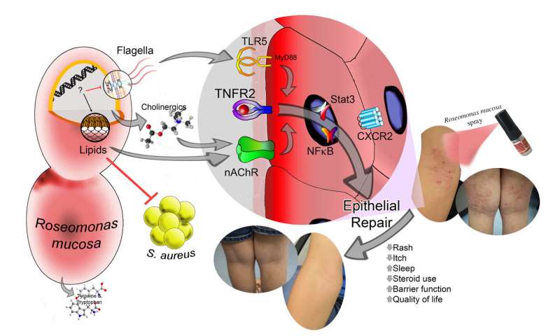
Topical application of Roseomonas mucosa was associated with clinical improvement of atopic dermatitis in 15 children. The improvement may be related to lipid production by the bacteria which influence TNFR2-mediated epithelial repair through influences on nicotinic acetylcholine signaling and flagellar recognition. Credit: Art by Ian Myles

Twice weekly for three months and every other day for an additional month, children or their caregivers sprayed a solution of sugar water containing live R. mucosa onto areas of skin with eczema. For the first 15 children enrolled in the study, the dose of live R. mucosa was gradually increased each month. The last five children to enroll received the same dose throughout the four-month treatment period. Regardless of dosing strategy, no serious adverse events were attributed to the therapy.

"Most children in the study experienced substantial improvements in their skin and overall wellbeing following R. mucosa therapy. Encouragingly, the therapeutic bacteria stayed on the skin and continued to provide benefit after therapy stopped," said NIAID's Ian Myles, M.D., principal investigator of the trial. "These results support a larger study to further assess the safety and effectiveness of this  by comparing it with a placebo."

Seventeen of the 20 children experienced a greater than 50% improvement in eczema severity following treatment. Improvement occurred on all treated skin sites, including the inner elbows, inner knees, hands, trunk, and neck. The scientists also observed increases in the skin's barrier function—its ability to seal in moisture and keep out allergens. Additionally, most children needed fewer corticosteroids to manage their eczema, experienced less itching, and reported a better quality of life following the therapy. These benefits persisted after treatment ended, and the therapeutic R. mucosa strains remained on the skin for up to eight months.

The NIAID researchers next set out to better understand how R. mucosa therapy improves eczema symptoms. They found that treated skin had increased microbial diversity and reduced levels of Staphylococcus aureus—a bacterium known to exacerbate eczema.

In addition to imbalances in the microbiome, the skin of people with eczema is deficient in certain lipids, or oils. By conducting experiments in cell and animal models of eczema, the NIAID scientists found that a specific set of lipids produced by R. mucosa strains isolated from healthy skin can induce skin repair processes and promote turnover of skin tissue. Study participants had increased levels of these lipids on their  after treatment with R. mucosa.

The researchers emphasize that additional studies are needed to further elucidate the mechanism of R. mucosa therapy and to explore whether genetic or other factors may explain why some participants did not benefit from the experimental treatment. (by )


Source: https://medicalxpress.com/news/2020-09-probiotic-skin-therapy-eczema-children.html 


Robust and widespread antibody testing has emerged as a key strategy in the fight against SARS-CoV-2, the virus responsible for the COVID-19 pandemic. However current testing methods are too inaccurate or too expensive to be feasible on a global scale. But now, scientists at the Okinawa Institute of Science and Technology Graduate University (OIST) have developed a rapid, reliable, and low-cost antibody test.

The device, described in a proof-of-concept study published this week in Biosensors and Bioelectronics, uses portable lab-on-a-chip technology to accurately measure the concentration of antibodies present in diluted blood plasma.

Antibodies are proteins produced by the immune system to neutralize the virus. Research has found that COVID-19 antibodies are present in the later stages of infection and can linger in the blood after the infection has cleared, allowing previously infected individuals to be identified. Antibody tests are thus an important means of determining the full spread of the coronavirus -- information that is crucial to guide public health policies.

And yet many nations have so far failed to employ large-scale antibody testing.

"Many existing platforms for antibody tests are accurate and reliable, but they are costly and need to be carried out in a lab by trained operators. This means that it can take hours, or even days, to obtain results," said Dr. Riccardo Funari, first author and a postdoctoral researcher in the Micro/Bio/Nanofluidics Unit at OIST. "Other tests are easier to use, portable and rapid, but are not sufficiently accurate, which hampers testing efforts."

The researchers avoided this trade-off between accuracy and accessibility by developing an alternative antibody testing platform that combines powerful light-sensing technology with a microfluidic chip. The chip provides results within 30 minutes and is highly sensitive, detecting even the lowest clinically-relevant antibody concentration. Each chip is cheap to manufacture and negates the need for a lab or trained operators, increasing the feasibility of nation-wide testing.

And there's another distinctive advantage of this newly developed platform. "The test doesn't just detect whether the antibodies are present or absent -- it also provides information about the quantity of antibodies produced by the immune system. In other words, it's quantitative," said Professor Amy Shen, who leads the Micro/Bio/Nanofluidics Unit. "This greatly expands its potential applications, from treating COVID-19 to use in developing vaccines."

Illuminating the antibodies

The antibody testing platform consists of a microfluidic chip which is integrated with a fiber-optic light probe. The chip itself is made from a gold-covered glass slide with an embedded microfluidic channel. Using an electric voltage, the team fabricated tens of thousands of tiny spiky gold structures, each one smaller than the wavelength of light, on a glass slide.

The researchers then modified these gold nanospikes by attaching a fragment of the SARS-CoV-2 spike protein. This protein is crucial for helping the coronavirus infect cells and causes a strong reaction from an infected person's immune system.

In this proof-of-concept study, the scientists demonstrated the principle behind how the test detects antibodies by using artificial human plasma sample spiked with COVID-19 antibodies that are specific to the spike protein.

Using a syringe pump, the sample is drawn through the chip. As the plasma flows past the protein-coated gold nanospikes, the antibodies bind to the spike protein fragments. This binding event is then detected by the fiber optic light probe.

"The detection principle is simple but powerful," said Dr. Funari. He explained that is it based on the unique behavior of electrons on the surface of the gold nanospikes, which oscillate together when hit by light. These resonating electrons are highly sensitive to changes in the surrounding environment, such as the binding of antibodies, which causes a shift in the wavelength of light absorbed by the nanospikes.

"The more antibodies that bind, the larger the shift in the wavelength of the absorbed light," added Dr. Funari. "The fiber optic probe is connected to a light detector which measures this shift. Using that information, we can determine the concentration of antibodies within the plasma sample."

A bright future

The large-scale roll-out of a quantitative test could greatly impact how COVID-19 is treated.

For example, quantitative tests could help doctors track how effectively a patient's immune system is fighting the virus. It could also be used to help identify suitable donors for a promising experimental treatment, called plasma transfusion therapy, where a recovered patient's antibody-rich blood is donated to currently infected patients to help them fight the virus.

Being able to measure the level of immune response can also aid vaccine development, allowing researchers to determine how effectively a trial vaccine triggers the immune system.

However, the researchers emphasized that the device is still undergoing active development. The unit aims to reduce the chip size to cut manufacturing costs and is also working on improving the reliability of the test.

"We have shown that the device works to detect different concentrations of the spike protein antibody in artificial human plasma samples. We now want to expand the test so that the chip can detect multiple different antibodies at the same time," said Dr. Funari. "Once the device is optimized, we plan to collaborate with local hospitals and medical institutions to perform tests on real patient samples."


Story Source:

Materials provided by Okinawa Institute of Science and Technology (OIST) Graduate University. Originally written by Dani Ellenby. Note: Content may be edited for style and length.


Journal Reference:

  1. Riccardo Funari, Kang-Yu Chu, Amy Q. Shen. Detection of antibodies against SARS-CoV-2 spike protein by gold nanospikes in an opto-microfluidic chipBiosensors and Bioelectronics, 2020; 169: 112578 DOI: 10.1016/j.bios.2020.112578


Source:https://www.sciencedaily.com/releases/2020/09/200908131038.htm 

Subcategories

Featured Links

PNHRS

http://www.healthresearch.ph

PCHRD

http://www.pchrd.dost.gov.ph

eHealth

http://www.ehealth.ph

Ethics

http://ethics.healthresearch.ph

ASEAN-NDI

http://www.asean-ndi.org

Events Calendar

January 2025
S M T W T F S
29 30 31 1 2 3 4
5 6 7 8 9 10 11
12 13 14 15 16 17 18
19 20 21 22 23 24 25
26 27 28 29 30 31 1