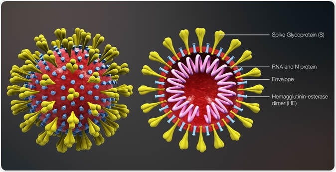
Robust and widespread antibody testing has emerged as a key strategy in the fight against SARS-CoV-2, the virus responsible for the COVID-19 pandemic. However current testing methods are too inaccurate or too expensive to be feasible on a global scale. But now, scientists at the Okinawa Institute of Science and Technology Graduate University (OIST) have developed a rapid, reliable, and low-cost antibody test.

The device, described in a proof-of-concept study published this week in Biosensors and Bioelectronics, uses portable lab-on-a-chip technology to accurately measure the concentration of antibodies present in diluted blood plasma.

Antibodies are proteins produced by the immune system to neutralize the virus. Research has found that COVID-19 antibodies are present in the later stages of infection and can linger in the blood after the infection has cleared, allowing previously infected individuals to be identified. Antibody tests are thus an important means of determining the full spread of the coronavirus -- information that is crucial to guide public health policies.

And yet many nations have so far failed to employ large-scale antibody testing.

"Many existing platforms for antibody tests are accurate and reliable, but they are costly and need to be carried out in a lab by trained operators. This means that it can take hours, or even days, to obtain results," said Dr. Riccardo Funari, first author and a postdoctoral researcher in the Micro/Bio/Nanofluidics Unit at OIST. "Other tests are easier to use, portable and rapid, but are not sufficiently accurate, which hampers testing efforts."

The researchers avoided this trade-off between accuracy and accessibility by developing an alternative antibody testing platform that combines powerful light-sensing technology with a microfluidic chip. The chip provides results within 30 minutes and is highly sensitive, detecting even the lowest clinically-relevant antibody concentration. Each chip is cheap to manufacture and negates the need for a lab or trained operators, increasing the feasibility of nation-wide testing.

And there's another distinctive advantage of this newly developed platform. "The test doesn't just detect whether the antibodies are present or absent -- it also provides information about the quantity of antibodies produced by the immune system. In other words, it's quantitative," said Professor Amy Shen, who leads the Micro/Bio/Nanofluidics Unit. "This greatly expands its potential applications, from treating COVID-19 to use in developing vaccines."

Illuminating the antibodies

The antibody testing platform consists of a microfluidic chip which is integrated with a fiber-optic light probe. The chip itself is made from a gold-covered glass slide with an embedded microfluidic channel. Using an electric voltage, the team fabricated tens of thousands of tiny spiky gold structures, each one smaller than the wavelength of light, on a glass slide.

The researchers then modified these gold nanospikes by attaching a fragment of the SARS-CoV-2 spike protein. This protein is crucial for helping the coronavirus infect cells and causes a strong reaction from an infected person's immune system.

In this proof-of-concept study, the scientists demonstrated the principle behind how the test detects antibodies by using artificial human plasma sample spiked with COVID-19 antibodies that are specific to the spike protein.

Using a syringe pump, the sample is drawn through the chip. As the plasma flows past the protein-coated gold nanospikes, the antibodies bind to the spike protein fragments. This binding event is then detected by the fiber optic light probe.

"The detection principle is simple but powerful," said Dr. Funari. He explained that is it based on the unique behavior of electrons on the surface of the gold nanospikes, which oscillate together when hit by light. These resonating electrons are highly sensitive to changes in the surrounding environment, such as the binding of antibodies, which causes a shift in the wavelength of light absorbed by the nanospikes.

"The more antibodies that bind, the larger the shift in the wavelength of the absorbed light," added Dr. Funari. "The fiber optic probe is connected to a light detector which measures this shift. Using that information, we can determine the concentration of antibodies within the plasma sample."

A bright future

The large-scale roll-out of a quantitative test could greatly impact how COVID-19 is treated.

For example, quantitative tests could help doctors track how effectively a patient's immune system is fighting the virus. It could also be used to help identify suitable donors for a promising experimental treatment, called plasma transfusion therapy, where a recovered patient's antibody-rich blood is donated to currently infected patients to help them fight the virus.

Being able to measure the level of immune response can also aid vaccine development, allowing researchers to determine how effectively a trial vaccine triggers the immune system.

However, the researchers emphasized that the device is still undergoing active development. The unit aims to reduce the chip size to cut manufacturing costs and is also working on improving the reliability of the test.

"We have shown that the device works to detect different concentrations of the spike protein antibody in artificial human plasma samples. We now want to expand the test so that the chip can detect multiple different antibodies at the same time," said Dr. Funari. "Once the device is optimized, we plan to collaborate with local hospitals and medical institutions to perform tests on real patient samples."


Story Source:

Materials provided by Okinawa Institute of Science and Technology (OIST) Graduate University. Originally written by Dani Ellenby. Note: Content may be edited for style and length.


Journal Reference:

  1. Riccardo Funari, Kang-Yu Chu, Amy Q. Shen. Detection of antibodies against SARS-CoV-2 spike protein by gold nanospikes in an opto-microfluidic chipBiosensors and Bioelectronics, 2020; 169: 112578 DOI: 10.1016/j.bios.2020.112578


Source:https://www.sciencedaily.com/releases/2020/09/200908131038.htm 

Hundreds of innovators, research pioneers, clinicians, industry leaders, and policymakers from all around Europe are united by a vision of how to revolutionize healthcare. In two publications—a perspective article in the journal Nature and the LifeTime Strategic Research Agenda—they now present a detailed roadmap of how to leverage the latest scientific breakthroughs and technologies over the next decade to track, understand, and treat human cells throughout an individual's lifetime.

The LifeTime initiative, co-coordinated by the Max Delbrueck Center of Molecular Medicine in the Helmholtz Association (MDC) in Berlin and the Institut Curie in Paris, has developed a strategy to advance personalized treatment for five major disease classes: cancer, neurological, infectious, chronic inflammatory and cardiovascular diseases. The aim is a new age of personalized, cell-based interceptive medicine for Europe with the potential of improved health outcomes and more cost-effective treatment, resulting in profoundly changing a person's healthcare experience.

Earlier detection and more effective treatment of diseases

To form a functioning, healthy body, cells follow developmental paths during which they acquire specific roles in tissues and organs. But when they deviate from their healthy course, they accumulate changes leading to disease which remain undetected until symptoms appear. At this point,  is often invasive, expensive, and inefficient. However, now we have the technologies to capture the molecular makeup of individual cells and to detect the emergence of disease or therapy resistance much earlier.

Using breakthrough single-cell and imaging technologies in combination with artificial intelligence and personalized disease models will allow us to not only predict disease onset earlier but also to select the most effective therapies for individual patients. Targeting disease-causing cells to intercept disorders before irreparable damage occurs will substantially improve the outlook for many patients and has the potential of saving billions of Euros of disease-related costs in Europe.

A detailed roadmap for implementing LifeTime

The perspective article, titled "The LifeTime initiative and the future of cell-based interceptive medicine in Europe," and the LifeTime Strategic Research Agenda (SRA) explain how these technologies should be rapidly co-developed, transitioned into clinical settings, and applied to the five major disease areas. Close interactions between European infrastructures, , hospitals, and industry will be essential to generate, share, and analyze LifeTime's big medical data across European borders. The initiative's vision advocates ethically responsible research to benefit citizens all across Europe.

According to Professor Nikolaus Rajewsky, scientific director of the Berlin Institute for Medical System Biology at the Max Delbrueck Center for Molecular Medicine and coordinator of the LifeTime Initiative, the LifeTime approach is the way into the future: "LifeTime has brought together scientists across fields—from biologists to clinicians, data scientists, engineers, mathematicians, and physicists ¬- to enable a much-improved understanding of molecular mechanisms driving health and disease. Cell-based medicine will allow doctors to diagnose diseases earlier and intercept disorders before irreparable damage has occurred. LifeTime has a unique value proposition that promises to improve the European patient's health."

Dr. Geneviève Almouzni, director of research at CNRS, honorary director of the research center from Institut Curie in Paris, and co-coordinator of the LifeTime Initiative believes that the future with LifeTime offers major social and : "By implementing interceptive, cell-based medicine we will be able to considerably improve treatment across many diseases. Patients all over the world will be able to lead longer, healthier lives. The economic impact could be tremendous with billions of Euros saved from productivity gains simply for cancer, and significantly shortened ICU stays for COVID-19. We hope EU leaders will realize we have to invest in the necessary research now." (by 

Source: https://medicalxpress.com/news/2020-09-health-cell-based-interceptive-medicine.html 

Researchers at Karolinska Institutet in Sweden have identified a small neutralizing antibody, a so-called nanobody, that has the capacity to block SARS-CoV-2 from entering human cells.

The researchers believe this nanobody has the potential to be developed as an antiviral treatment against COVID-19. The results are published in the journal Nature Communications.

"We hope our findings can contribute to the amelioration of the COVID-19 pandemic by encouraging further examination of this nanobody as a therapeutic candidate against this viral infection."

Gerald McInerney, Study Corresponding Author and Associate Professor of Virology, Department of Microbiology, Tumor and Cell Biology, Karolinska Institute

The search for effective nanobodies--which are fragments of antibodies that occur naturally in camelids and can be adapted for humans--began in February when an alpaca was injected with the new coronavirus' spike protein, which is used to enter our cells.

After 60 days, blood samples from the alpaca showed a strong immune response against the spike protein.

Next, the researchers cloned, enriched, and analysed nanobody sequences from the alpaca's B cells, a type of white blood cell, to determine which nanobodies were best suited for further evaluation.

They identified one, Ty1 (named after the alpaca Tyson), that efficiently neutralizes the virus by attaching itself to the part of the spike protein that binds to the receptor ACE2, which is used by SARS-CoV-2 to infect cells. This blocks the virus from slipping into the cells and thus prevents infection.

"Using cryo-electron microscopy, we were able to see how the nanobody binds to the viral spike at an epitope which overlaps with the cellular receptor ACE2-binding site, providing a structural understanding for the potent neutralisation activity."

Leo Hanke, Study First Author and Postdoc in the McInerney Group

Nanobodies offer several advantages over conventional antibodies as candidates for specific therapies. They span less than one-tenth the size of conventional antibodies and are typically easier to produce cost-effectively at scale.

Critically, they can be adapted for humans with current protocols and have a proven record of inhibiting viral respiratory infections.

"Our results show that Ty1 can bind potently to the SARS-CoV-2 spike protein and neutralize the virus, with no detectable off-target activity" says Ben Murrell, assistant professor in the Department of Microbiology, Tumor and Cell Biology and co-senior author of the publication. "We are now embarking on preclinical animal studies to investigate the neutralizing activity and therapeutic potential of Ty1 in vivo".

This project is the first arising from the CoroNAb consortium, which is coordinated by Karolinska Institutet, and funded by the European Union's Horizon 2020 research and innovation programme. Additional funding for this project was obtained from the Swedish Research Council and KI Development Office.

The sequence of Ty1 is available in the scientific article and will also be posted on the NCBI GenBank sequence database under the accession code MT784731.

 
Journal reference:

Ziegler, C. G. K., et al. (2020) SARS-CoV-2 Receptor ACE2 Is an Interferon-Stimulated Gene in Human Airway Epithelial Cells and Is Detected in Specific Cell Subsets across Tissues. Celldoi.org/10.1016/j.cell.2020.04.035.


Source: https://www.news-medical.net/news/20200904/Study-Small-neutralizing-antibody-can-block-SARS-CoV-2-from-entering-human-cells.aspx 

As the COVID-19 pandemic progresses, it is crucial to track the viral changes in order to link the clinical and epidemiological features of the disease with these mutations. A new study published on the preprint server bioRxiv* in September 2020 reports on a common severe acute respiratory syndrome coronavirus 2 (SARS-CoV-2) mutation, D614G, and its effects on the virus.

Early in the course of the pandemic, a new mutation emerged, namely, D614G, in the spike protein, superseding most local strains wherever it appeared. The current study is based on incorporating this mutation into a wildtype SARS-CoV-2 strain in order to understand how it affects virus-host interactions.

The outcomes they measured included viral replication on human lung epithelial cells and primary human airway tissues, viral fitness in hamster upper airway, and neutralization susceptibility. This study should contribute to understanding the role played by this mutation in the transmission of the virus, the effectiveness of various vaccines, and of convalescent antibody-rich plasma.

Spike Mutations and Their Importance

Even though most COVID-19 cases develop mild illness only, the pandemic has claimed almost 900,000 lives worldwide in the last eight months. Notably, severe COVID-19 is linked to dysregulated immune response, with excessive and systemic inflammation. However, viral factors such as virulence could also play a part in the evolution of the illness.

Researchers have discovered mutations that encode amino acids of a different sort in the SARS-CoV-2 spike protein sequence. This protein is key for viral entry into the host cell via the angiotensin-converting enzyme 2 (ACE2). Such mutations may cause alterations in the host range, pathogenesis and tissue tropism.

In the earlier SARS outbreak of 2002, a single mutation in the spike protein allowed the infection to spill over from the original reservoir to the intermediate civet host and to humans.

The D614G Mutation

In the current pandemic, the D614G mutation has become dominant after March, to cover ~75% of all published viral sequences by June 2020. There were three accompanying mutations as well.

This mutation set has become the predominant one both worldwide and also within distinct localities after introduction, showing that it may have conferred a fitness advantage and not just due to the founder effect or genetic drift. One especial advantage appears to be increased infectious potential, supported by the finding that this mutant is associated with higher viral loads in the nasopharynx in COVID-19 patients.

Pseudoviruses Show Improved Infectivity of Mutant Strain

Transmissibility is a key factor in viral fitness, but direct measurements were required to confirm the role of the mutation in improved fitness. The researchers first described the phenotype of the D614G mutation in pseudoviruses. They found higher viral titers in many different types of cell cultures when infected by the G614 variant. This indicated that perhaps the variant could cause higher viral entry into cells and replication in the airway.

To confirm this, they used wildtype virus with the spike mutation to infect a cell culture, primary human airway 3D tissue, and experimental hamsters. They also used neutralization tests for serum samples and monoclonal antibodies (mAbs), based on reporter SARS-CoV-2 viruses carrying either D614 or G614, labeled with mNeonGreen.

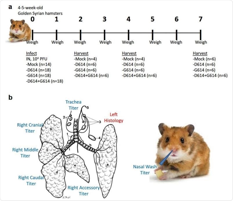
Experimental design of hamster infection and sample harvest. (a) Graphical overview of experiment to assess the impact of G614 mutation on replication in the respiratory system of hamsters. (b) Schematic samples harvested on days 2, 4, and 7 post-infection. Illustration of hamster lung adapted from Reznik, G. et al. Clinical anatomy of the European hamster. Cricetus cricetus, L.

Higher Replication and Infectivity of Mutant Strain

The researchers observed that the D614G substitution in the spike protein in human lung epithelial cells caused more significant viral replication and infectivity by comparing the two variants, first in Vero cell culture. They found similar-looking plaques, but with a higher infectious titer with the G614 virus after 12 hours; later, both displayed similar titers. The same was seen for extracellular viral RNA loads. This showed that the mutation did not affect these two parameters on Vero cells.

The next step was to repeat the experiment on human lung epithelial Calu3 cells. They found that the G614 virus had somewhat higher infectious viral titers up to a maximum of 2.4 times higher over the next 48 hours. Still, the viral RNA load was lower or equivalent over the same period with this variant. This indicates that the D614G mutation makes the virus in the human lung cell line more infectious.

How did this happen? To know more, the researchers then looked at how the spike protein was being processed in the two variants. They found that full-length spike protein obtained from the virions grown in human lung epithelial cells was almost fully processed to the cleavage form in both variants. However, those obtained from Vero cells showed markedly lower cleavage efficiencies by 20% and 30% less for the D614 and G614, respectively.

This suggests the influence of the cell culture on the cleavage efficiency, but no differential effect was observed between the two viruses.

Mutation Improves Fitness in Hamster Upper Airway

The D614G mutation was then tested in the golden Syrian hamster model, where it was found that either variant showed similar pathological features, including weight loss, but without other visible symptoms. The infectious viral titers from the upper and lower respiratory tract were similar for both viruses on the second day, but the differences were more apparent in the upper than the lower airway. The most significant difference between the titer of infectious virus for the two variants was seen on day 4 after infection, in the upper airway, particularly in the nasal epithelium.

Even though the infectious viral titer was higher for G614 than for D614 in hamsters, the viral RNA load was almost identical. Thus, the ratio of RNA to PFU for G614 remained lower across the range of airway tissues.

The researchers' comment, “If the lower viral RNA/PFU ratio of the G614 virus could be extrapolated to COVID-19 patients, the modest differences in cycle threshold (Ct) values of RT-qPCR in patients’ nasopharyngeal swabs would translate to ≥10-fold infectious G614 virus, underscoring the potential for enhanced transmission and spread.” In fact, it has been observed that if the same patient has two different strains, one in throat swabs and the other in sputum, the former alone is the cause of spread.

On the seventh day after infection, no infectious virus could be detected. However, viral RNA copies were still abundantly found in nasal wash samples, indicating that viral RNA persists after the infectious virions are cleared. This explains positive RT-PCR findings in some COVID-19 patients, even when the infectious virus is undetectable. Moreover, it reflects the clinical finding that disease severity is not correlated with the dominant mutation.

Direct fitness comparison was carried out using a competition experiment. This allows the elimination of host-host variations while using internal controls, with more precise quantification of the ratio of the virus strains than comes from finding the titers individually. Using intranasal inoculation, they infected hamsters with 104 PFU each for each virus. To compensate for the Vero cells of origin, they were also given viral RNA to levels equivalent to the above. They found that in all airway tissues, the G614 virus is superior to D614G614/D614, indicating its replication advantage.

Viral Replication Boosted

The researchers then evaluated the replication of D614 and G614 viruses in a 3D primary airway tissue model. They found that titers of infectious virus were much higher for the latter, at 2-8 times, but not the viral RNA loads. This confirms the effect of the substitution on replication by increasing viral replication in the human upper airway tissue.

Competition experiments to compare replication fitness showed that even when the initial infectious ratio of D614 and G614 went from 1:1 to 9:1, the latter still increased to fivefold the infectious viral titer of the former within five days. The G614 strain can thus outperform the other at great speed to achieve the numerical advantage, even when it is first present as the minor variant in a mixed virus population.

Susceptibility to Neutralization

The neutralization testing of a set of sera collected from hamsters that had been infected with the D614 strain showed that in all cases, the neutralization titer required for G614 was 1.4- to 2.3 times higher than for D614, showing that this mutation may increase the susceptibility to neutralization. Thus, any vaccine effective against the former will not lose its efficacy against the latter.

The researchers then looked at 11 anti-RBD mAbs against the two viruses. They found that only one showed double the potency against G614 compared to D614, but the other 10 showed similar neutralization efficacy to both. The mutation may reduce the neutralization efficacy depending on the conformation of the spike protein, based on which epitope is presented to the mAb.

Conclusion

The researcher sum up their findings as demonstrating that “spike substitution D614G enhances viral replication in the upper respiratory tract and increases neutralization susceptibility.” This could help develop better vaccines and antibodies to contain the pandemic in the future. (written By Dr. Liji Thomas, MD)

*Important Notice

bioRxiv publishes preliminary scientific reports that are not peer-reviewed and, therefore, should not be regarded as conclusive, guide clinical practice/health-related behavior, or treated as established information.

Journal reference:

In a ceremonial turnover on Friday, 4 September 2020, the Department of Health (DOH) will formally receive the Feasibility Analysis of Syndromic Surveillance Using Spatio-Temporal Epidemiological Modeler For Early Detection of Diseases, commonly called FASSSTER, as a tool to aid in disease surveillance in the country, including the monitoring of COVID-19.

The disease surveillance tool (FASSSTER) was developed by the Ateneo De Manila University (ADMU), with funding grant from the Department of Science and Technology - Philippine Council for Health Research and Development (DOST-PCHRD), in 2016, initially for dengue, but which has since been recalibrated as a web-based disease surveillance platform that allows policymakers to understand the outbreaks at the national, regional, and local levels and assess the effects of the preventive measures in place. 

As a disease surveillance tool for COVID-19, the Ateneo Center for Computing Competency and Research (ACCCRe) of ADMU collaborated with the University of the Philippines Manila - National Telehealth Center (UP-NTHC) and the Department of Health-Epidemiology Bureau to develop the technology which is now publicly accessible at https://fassster.ehealth.ph/covid19 and uses localized indices from Philippine health records.

It is indeed heartwarming to see our experts in FASSSTER actively contributing to public health through research and innovation. I share the pride with our researchers in saying that what started as a modest project in data science has today become a vital support to our healthcare system, especially in these uncertain times,” said DOST-PCHRD Executive Director Dr. Jaime Montoya.

The Council invites the public to attend the ceremonial turnover by registering through this link. Source: http://www.pchrd.dost.gov.ph/index.php/news/6596-doh-adopts-dost-ateneo-fassster-disease-surveillance-tool (Written by: Christine Jane Gonzalez)

Subcategories

Featured Links

PNHRS

http://www.healthresearch.ph

PCHRD

http://www.pchrd.dost.gov.ph

eHealth

http://www.ehealth.ph

Ethics

http://ethics.healthresearch.ph

ASEAN-NDI

http://www.asean-ndi.org

Events Calendar

April 2025
S M T W T F S
30 31 1 2 3 4 5
6 7 8 9 10 11 12
13 14 15 16 17 18 19
20 21 22 23 24 25 26
27 28 29 30 1 2 3